During my deployment at Bagram Air Field, Afghanistan, I saw firsthand the ramifications associated with a lack of treatment options for skull repair in developing countries.
In one particular case, a young boy was severely injured after reaching to pick up what he thought was a toy buried in the ground. It turned out to be one of millions of land mines left by the Soviet Union during withdrawal from the Soviet-Afghan war. The mine exploded, and the young boy needed a decompressive craniectomy performed by military neurosurgeons to relieve pressure in the skull and save his life. He later returned for a cranioplasty, a procedure that restores the integrity of the skull with an implant. In this case, titanium mesh was used—a common option in countries with limited resources. Unfortunately, this material is known to be associated with an increased risk of delayed wound complications. By the time I saw the patient, the titanium mesh had eroded through his skin. He needed several additional procedures to repair the damage.
Cases like this are typical in developing countries. When patients acquire a bone defect, it is preferable in most cases to use the patient’s own bone flap. When the flap is not salvageable, synthetic materials are the next best option. Here in the United States, third-party companies produce anatomically precise patient-specific cranial implants using the patient’s CT scans. However, the cost for this type of implant is often prohibitive in countries with developing economies. Options for calvarial reconstruction in these regions are limited in many regards. Titanium mesh implants do not readily conform to many three-dimensional defects and can be associated with wound contracture, poor cosmesis (cosmetic appearance) and delayed wound erosion. Polymethyl methacrylate (PMMA), or bone cement, implants have to be manually shaped or directly poured into the defect. The cosmesis of this form of implant is often limited and depends on the ability of the surgeon to shape the implant appropriately before the cement hardens.
Developing a More Affordable Option
Is there a better option available? Researchers at UC believe that, with utilization of additive manufacturing or 3D printing, a feasible solution may exist.
In recent years, 3D printing has become more commonplace in medical research and personal use. An entry level 3D printer costs about one-quarter of the price of a single patient-specific cranial implant (PSCI) in the United States. Existing studies have demonstrated success with printing PSCIs and molds for cranioplasties. The postoperative cosmesis, in particular, has been excellent.
Why, then, has the solution not been widely utilized by surgeons in developing countries? The answer lies in the time commitment required to master the computer software necessary to transition from CT imaging to a .STL file of PSCI ready for printing. The lengthy training process and the necessary hours at the computer to generate each mold is more than enough to deter a busy surgeon.
With knowledge of this obstacle, I have partnered with Sam Anand, PhD, MS, professor of mechanical engineering at the UC College of Engineering and Applied Science, to research a way to simplify the process for surgeons in developing countries. The goal is to make this technology freely available to neurosurgeons around the world.
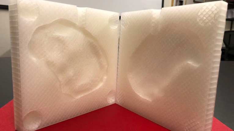
The aim of the project is to create a freeware program that allows neurosurgeons to upload CT scans of patients with a skull defect with near-instantenous generation of a .STL file ready for 3D printing. The program will be: (i) free, (ii) reproducible with minimal clinician training, and (iii) anatomically accurate for the defect. Dr. Anand’s research team has carefully constructed an algorithm for this research program with the needs of neurosurgeons in developing countries in mind. Printed implants using PMMA or titanium are the most convenient for intraoperative use but require more sophisticated printers and options for sterilization. The best option for surgeons in developing countries is a 3D printed mold that can be sterilized and allow casting of a PSCI in the operating room. Under sterile conditions in the OR, sterile bone cement can be injected into the mold and be ready for insertion in under 30 minutes.
How does this new software program rapidly generate a .STL file of a PSCI mold? The innovative concept behind the development of this software revolves around the utilization of the patient’s own skull as a template. The contralateral side of the skull across from the missing bone is mirrored onto the defect, which facilitates precise acquisition of flap margins. The Boolean difference is obtained, which generates a virtual implant. The virtual implant is then used to create a two-piece mold able to support intraoperative injection of bone cement. The final implant precisely restores the skull surface with symmetrical surface contours, ideal thickness and a seamless fit.
Making 3D Printed Cranioplasty a Reality
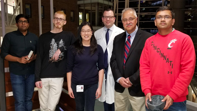
The Center for Global Design and Manufacturing at the University of Cincinnati is collaborating with us to turn this idea into reality. Dr. Anand, the center’s director, leads a team of engineers with extensive experience in additive manufacturing and software programming. Three graduate students, Omkar Ghalsasi (MS student) and Vysakh Venugopal and Matthew McConaha (PhD students) are working on this project under Dr. Anand’s supervision with Alice Xu, a third year medical student at the University of Cincinnati.