- After completing colorectal cancer treatment, Laura’s follow-up scan revealed a concerning lung nodule.
- Her University of Cincinnati Cancer Center team moved quickly, coordinating care across specialties and using minimally invasive lung surgery with advanced imaging technology.
- The tumor was fully removed — bringing clarity, confidence and a clear path forward.
Seeing Clearly and Moving Forward
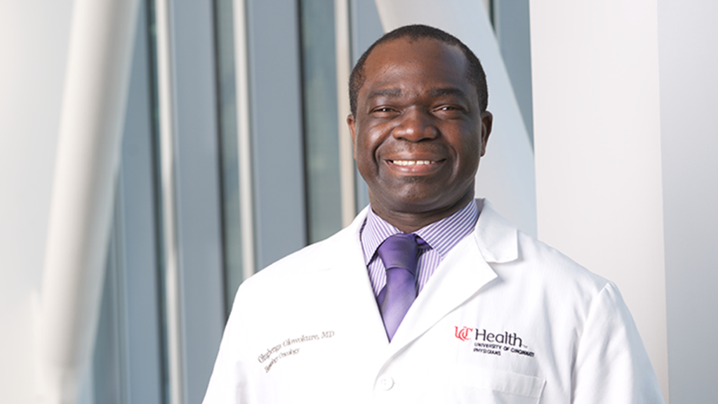
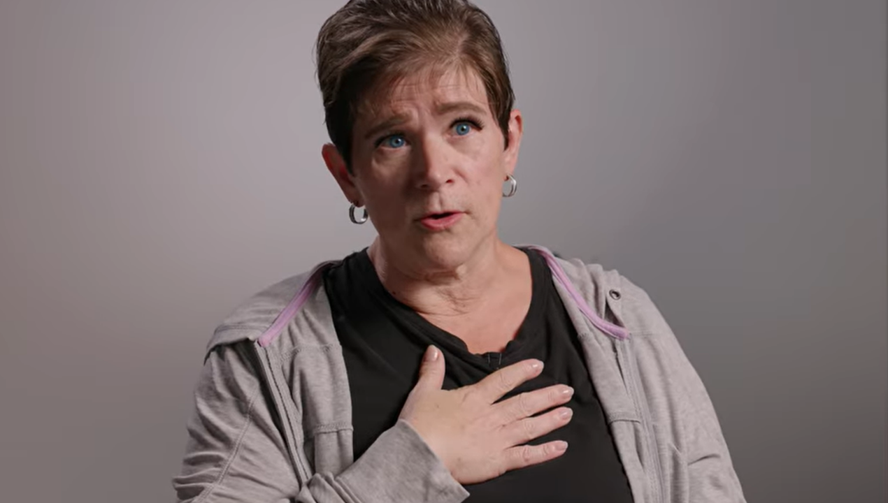
Laura Pearson had already been through chemotherapy, surgery and radiation for colorectal cancer under the care of Olugbenga Olowokure, MD, through UC Cancer Center’s Gastrointestinal Cancer Center. After everything she had endured, follow-up scans were supposed to offer reassurance. Instead, this one brought new questions.
A suspicious spot on the lung can be anxiety-producing, especially after years of cancer treatment and surveillance.
A lung nodule is a small spot or shadow seen on a scan. Many are harmless. But in someone with a history of cancer, even a small nodule can raise concern. Was this a new lung cancer? Or had her previous cancer spread?
“I didn’t want to wait,” she says. “I wanted certainty.”
Coordinated Care When It Matters Most
Because Laura was already a patient at the UC Cancer Center, her care moved quickly and seamlessly. Dr. Olugbenga partnered directly with Sandra Starnes, MD, Chief of Thoracic Surgery, to evaluate the next steps. Instead of navigating referrals on her own, Laura’s team coordinated her care across specialties — a benefit of comprehensive cancer treatment.
In cases like Laura’s, distinguishing between a new primary lung cancer and metastatic disease is critical. A primary lung cancer begins in the lung. A metastasis means cancer cells from another part of the body have traveled to the lung. The treatment approach, prognosis and follow-up plan can differ significantly.
A New Way to See During Surgery
Since Laura was found to be a good candidate, Dr. Starnes decided on an innovative surgical approach available at the UC Cancer Center.
Because the nodule’s exact nature could not be confirmed through imaging alone, surgery offered both diagnosis and treatment. Dr. Starnes performed a minimally invasive lung resection using video-assisted thoracoscopic surgery (VATS) — a technique that uses small incisions and a tiny camera to guide the procedure — allowing her to remove a small portion of lung tissue containing the tumor while preserving as much healthy lung as possible.
Instead of relying solely on pre-operative scans or tactile feedback in the operating room, Laura’s team used fluorescent imaging technology — a tool that helps surgeons see cancer more clearly during surgery.
Here’s how it works:
- A fluorescent agent is given intravenously before surgery.
- This agent binds to specific cells or structures and emits light when illuminated by a special imaging system.
- During the operation, areas that might otherwise be difficult to see light up — giving the surgeon clearer definition between healthy tissue and cancer in real time.
“It made it easier for the surgeon to see the definition of the tumor,” Laura says.
This real-time visualization provides added precision — helping ensure complete removal while minimizing unnecessary loss of healthy tissue. For patients, that can mean fewer complications, less pain and faster recovery.
Fast Turnaround, Clear Answers
During the procedure, Dr. Starnes removed a section of Laura’s lung, including the 1-centimeter tumor.
The result brought immense relief: the nodule was not a new primary lung cancer — it was a metastasis from her original colorectal cancer, and it was completely removed.
While the word “metastasis” can sound frightening, in Laura’s case the cancer was isolated and surgically treatable. Knowing exactly what it was allowed her team to tailor her follow-up care appropriately.
That clarity — knowing exactly what it was and that it was gone — was what Laura had hoped for.
“I was super happy about the fast turnaround,” she says. “Because not knowing… It’s just anxiety producing.”
Why This Technology Matters
Fluorescent imaging isn’t just a new tool — it represents a leap in surgical precision:
- Hidden Tumor Detection: Small tumors too subtle for the naked eye can light up during surgery.
- Clearer Tumor Margins: Surgeons can better distinguish diseased tissue from healthy tissue.
- Real-Time Guidance: Instead of relying on static pre-op images alone, surgeons see dynamic contrast as they operate.
- Improved Outcomes: Enhanced visualization can lead to more precise removal, fewer complications and potentially shorter recovery.
For patients with lung nodules — whether suspected lung cancer or metastatic disease — precision matters. Complete removal with preservation of healthy lung tissue supports long-term lung function and recovery.
For Laura, it meant certainty — not just about the surgery, but about what came after.
Healing After Surgery
Laura’s recovery from the surgery was quick — one night in the hospital and minimal pain — but the emotional impact was enduring.
“I feel well cared for now,” she says. “I feel very good about everything that’s gone on in the past and that will go on in the future.”
Today, Laura continues follow-up care with her oncology team. She continues to work and spend time with her grandkids, focusing not on uncertainty, but on living forward.
Her message for others:
“If something complicated is happening, surround yourself with a team that can listen, innovate, and act. Don’t settle for ambiguity.”
For Laura, the power of precision — and the ability to see clearly as a result of the surgery — brought the peace of mind she’d been seeking all along.
Why Choose University of Cincinnati Cancer Center
Through research and innovative techniques, clinicians at the University of Cincinnati Cancer Center a revolutionizing cancer care, from screening and early detection through follow up care.
- Top 20 Institutional Honor Roll – Thoracic Surgery Foundation
- U.S. News & World Report High-Performing Lung Cancer Surgery designation for six consecutive years
- Exclusive AI-assisted screening technology
For patients like Laura, that means answers without delay, precision without compromise and care that extends beyond a single specialty. Contact us directly at 513-585-UCCC (8222).