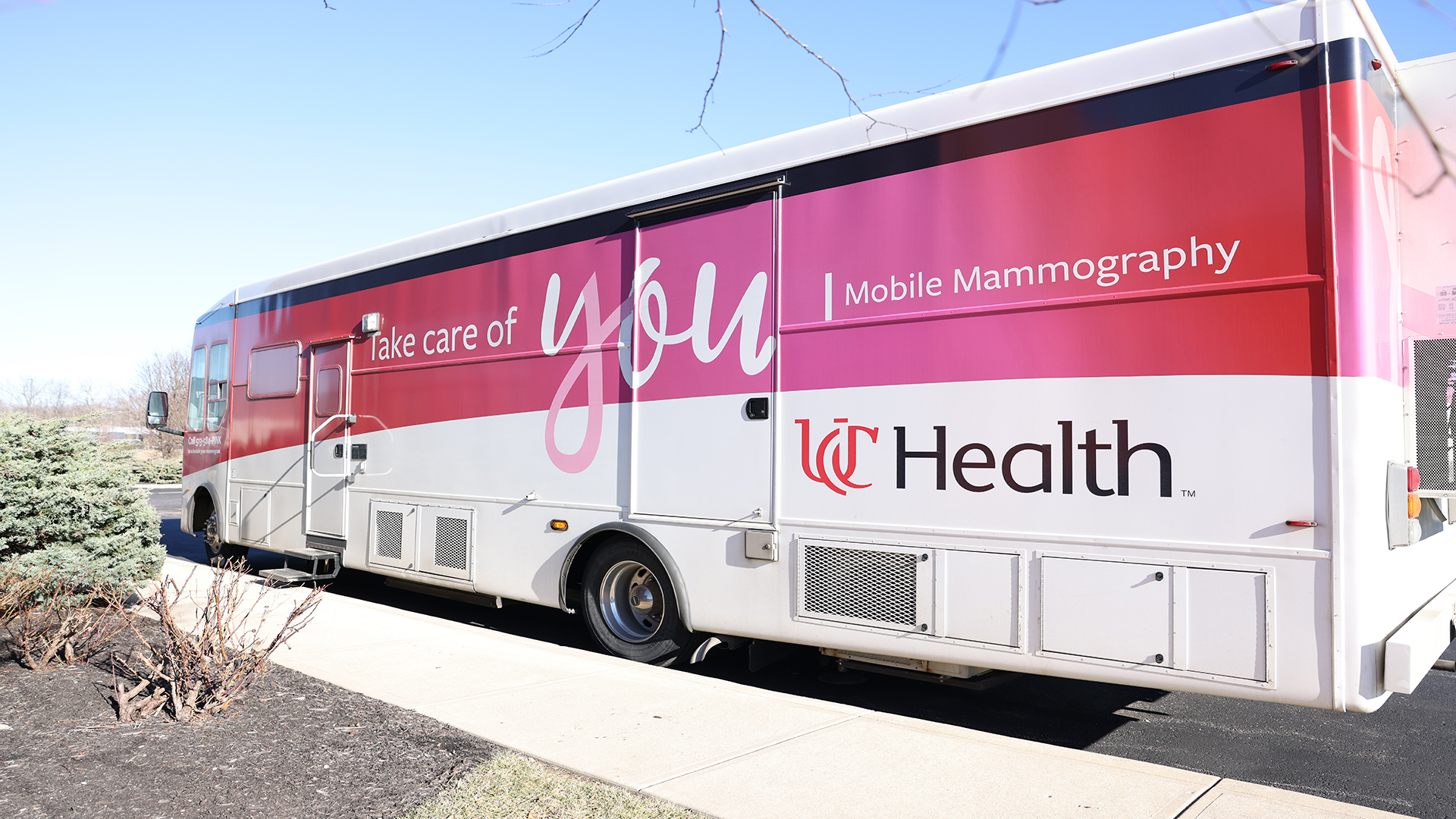
Your annual mammogram is an important part of your health. With flexible, comprehensive options, UC Health makes it easy to put your health first. Our mobile mammography unit travels throughout the city to provide convenient screening options, so you can access expert breast imaging close to home.
Radiology
Breast Imaging
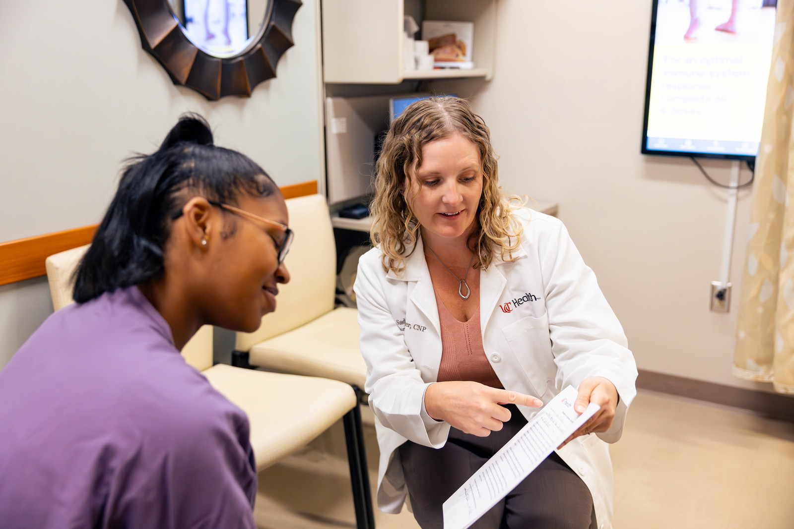
Personalized, expert breast screenings and diagnosis with UC Health’s compassionate team—using the latest technology and research to empower and support patients.
Breast Imaging
Innovation in Breast Imaging
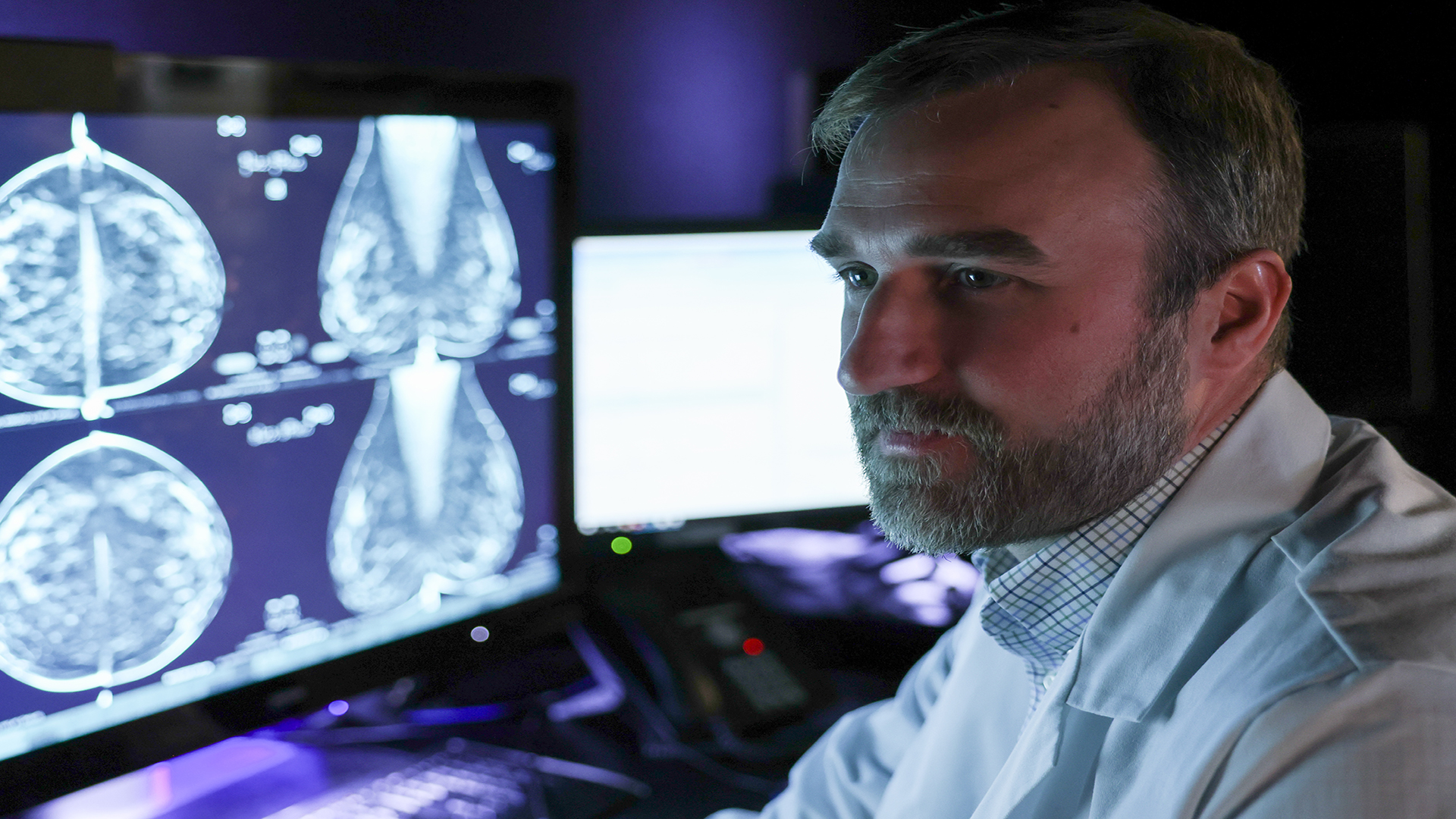
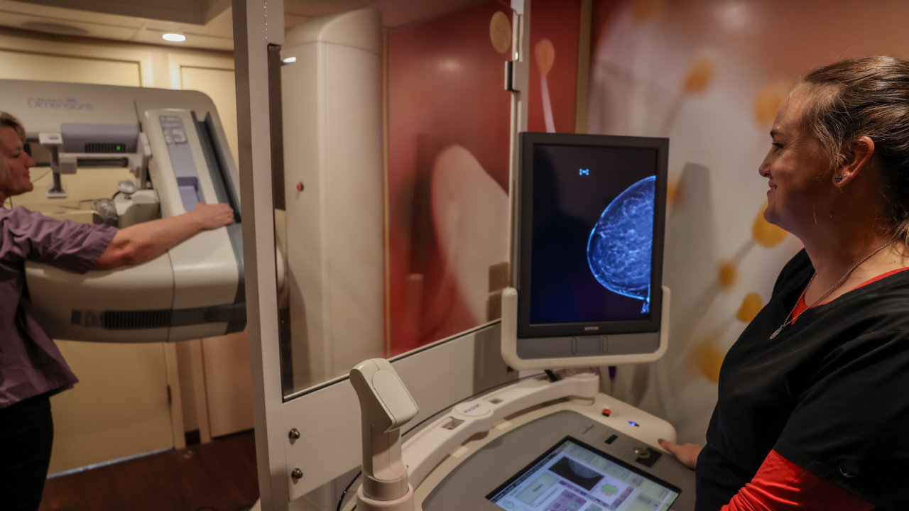
UC Health breast imaging radiologists continuously innovate and lead advancements in breast imaging technology. Our specialists consider your medical and family history to personalize your care, so you receive the most effective screening for your needs.
Breast Imaging
Double Accreditation in Breast Care
Our Breast Center has earned double accreditation for clinical excellence. Our dedicated and board-certified breast radiologists deliver proven expertise in every exam, ensuring you receive advanced, evidence-based care at every visit.
Breast Imaging
Research That Advances Care
Our team and patients contribute to research and clinical trials that enhance breast cancer screening and care. We are pioneering new studies, including ways to identify breast cancer before symptoms appear—giving you access to tomorrow’s care today.
Mobile Mammography
Research and Clinical Trials
Billing and Insurance
Mobile Mammography
Discover Convenient Ccreening Locations Close to You
Schedule your mammogram or find our mobile unit near you. Our team brings screening services throughout the city for easier access to lifesaving care.
Research and Clinical Trials
Explore Research Advancing Breast Cancer Detection
Learn how our team participates in research and clinical trials that improve breast cancer screening and care. See if you may be eligible to join a study.
Billing and Insurance
Get Answers on Billing, Insurance and Financial Support
Understand your medical bills and get help with payment options. Our team can guide you through insurance coverage and connect you with support resources.
Mobile Mammography
Mobile Mammography
Research and Clinical Trials
Research and Clinical Trials
Billing and Insurance
Billing and Insurance
Our Specialties
The Region’s Only Dedicated Team for Advanced Breast Imaging Care and Expertise
- Procedures We Offer
Search Results ({{searchSpecialtiesCount}})
{{ sortOrderLabel || 'Sort By' }}
Search Results ({{searchConditionsCount}})
{{ letter }}
- {{item.title}} {{item.title}}
{{ sortOrderLabel || 'Sort By' }}
Search Results ({{searchTreatmentsCount}})
- {{item.title}} {{item.title}}
Authoring Breakthroughs
National Leaders in Breast Imaging, Research and Personalized Care
Our Unique Approach
A Dedicated Team by Your Side
Our double-accredited Breast Center delivers the latest advances, collaboration and proven leadership so you feel supported, informed and confident—no matter where you are on your breast health journey.
2x
Our Breast Center is double accredited for excellence
Lead
Dedicated breast radiologists deliver proven expertise
Peace
Advanced screening technology provides accurate diagnosis
Options
Informing patients to make the best breast health choices
Collaborative, Personalized Care
Double-Accredited Breast Cancer Center
Our renowned breast oncologists, surgeons and radiologists collaborate to develop a personalized care plan just for you. Our Breast Center is a double-accredited center—meaning you always have access to the latest, evidence-based care.
Focused Expertise, Compassionate Care
Dedicated Breast Imaging for Every Patient
Our breast imaging experts specialize in breast screenings and diagnosis to ensure quality care. Our radiologists examine and interpret every detail of your breast tissue for precise evaluation. If an abnormality is detected, our team collaborates with breast health experts to determine your best care plan.


How to Contribute
Support Breast Imaging Innovation
Your donation advances medical research and breast care. Our research and treatments benefit patients in our community and worldwide, creating better breast imaging for everyone.

Contact Us
Contact Us
We are here to guide you with compassionate, expert breast imaging care.